TSUJI ORTHOPAEDIC INSTITUTE
PAINFUL LESION
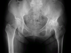
In this roentgenogram, the right side of the patient hip joint (left on the screen) is normal and the other side shows the typical x-ray appearance of the degenerative joint disease of the hip. Joint space is missing, the femoral head is oval (coxa planna), and the shaft of the femur shows osteoatrophy.